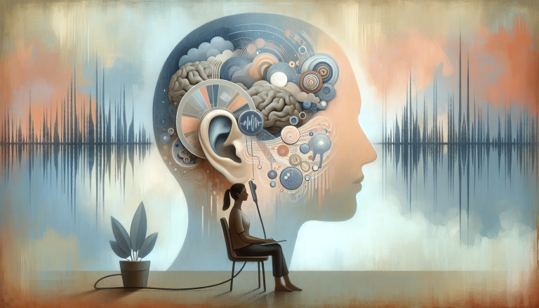
Tinnitus Phenotypes: Brain Connectivity and Activity Differences
Peer-Reviewed Research
Functional brain imaging has revealed distinct neural patterns between people who are troubled by their tinnitus and those who are not, according to a new case-control study. The research, led by Yongpeng Li, Lu Peng, and Ying Lan, compared 61 patients with bothersome tinnitus (BT), 52 with non-bothersome tinnitus (NBT), and 50 hospital-based controls without tinnitus using resting-state fMRI. Their analysis shows the brain’s functional architecture differs more between tinnitus phenotypes than between bothersome tinnitus and a non-tinnitus control group.
Key Takeaways
- Brain connectivity between the temporal lobes and the anterior cingulate/medial orbitofrontal cortex was stronger in people with bothersome tinnitus compared to those with non-bothersome tinnitus.
- Regional brain activity (fALFF and ReHo) showed consistent differences across multiple areas, most reliably distinguishing bothersome from non-bothersome tinnitus.
- After adjusting for perceived tinnitus loudness, many imaging differences between bothersome and non-bothersome groups were reduced, suggesting these neural patterns are linked to the clinical burden of the symptom.
- The brains of people with bothersome tinnitus did not show significant functional abnormalities compared to non-tinnitus controls, indicating these are phenotype-related features, not disease markers.
How the Brain Scan Study Was Conducted
The researchers recruited 163 participants, dividing them into three clear groups based on clinical assessment. All participants underwent resting-state functional MRI scans. The team analyzed the data using three established metrics: fractional amplitude of low-frequency fluctuations (fALFF) to measure regional spontaneous brain activity, regional homogeneity (ReHo) to assess local synchronization, and seed-based functional connectivity (FC). They used broad bilateral temporal lobe regions as seeds to examine connectivity within temporal-related networks, including links to limbic structures involved in emotion.
Group comparisons were performed using ANCOVA, controlling for a wide range of potential confounders including age, hearing thresholds, blood pressure, sleep quality, and emotional state. Statistical thresholds were set at a cluster-level false discovery rate (FDR) corrected p < 0.05. The researchers also conducted exploratory analyses to see if any imaging measures correlated with clinical scores for tinnitus distress, loudness, or hearing ability.
Connectivity and Activity Differences Define Tinnitus Distress
The imaging results painted a clear picture of divergence between the two tinnitus groups. A primary finding was a significant difference in functional connectivity between the bilateral temporal lobe seed and a cluster in the left anterior cingulate cortex (ACC) and medial orbitofrontal cortex (mOFC). Post-hoc tests showed this connectivity was stronger in the BT group than in the NBT group. Notably, the BT group’s connectivity did not differ from the hospital-based non-tinnitus controls.
Patterns of local brain activity also distinguished the groups. ReHo in the medial superior frontal gyrus was lower in the NBT group compared to both the BT group and controls. The fALFF analysis showed more widespread differences, with group variations observed in temporal, frontal, insular, occipital, supramarginal, and postcentral regions. The most consistent and reliable differences across these metrics were those observed between the BT and NBT groups.
These findings resonate with models of conditions like thalamocortical dysrhythmia, where altered neural rhythms are implicated in persistent perceptual disorders. The involvement of the ACC and mOFC—key nodes for emotional salience and evaluation—points to why the same auditory perception can be neutral for one person and deeply distressing for another.
Loudness and Burden, Not Just Presence, Drive Brain Changes
A critical part of the analysis tested the robustness of these findings. The researchers performed sensitivity analyses, keeping their original covariates but also adding a statistical adjustment for each participant’s subjective rating of tinnitus loudness on a visual analog scale (VAS). This adjustment attenuated most of the significant differences between the BT and NBT groups.
This result is a key interpretation. It suggests that the observed functional differences in connectivity and local activity are not simply a signature of tinnitus presence. Instead, they appear to be linked to the perceived intensity or the associated clinical burden of the condition. The brain’s functional architecture in bothersome tinnitus may reflect the brain’s engagement with a sound it deems salient and bothersome, rather than a static abnormality.
Furthermore, exploratory analyses found no significant correlations between the imaging indices and clinical measures like tinnitus handicap scores or pure-tone averages that survived FDR correction. This lack of direct correlation underscores the complexity of the relationship; the brain patterns are group-level features associated with the phenotype of “bother,” but they may not linearly track with individual symptom reports.
Implications for Understanding and Treatment
The study’s conclusion reframes how we might view the “tinnitus brain.” Because several core measures did not separate the bothersome tinnitus group from non-tinnitus controls, the authors caution against interpreting the findings as BT-specific abnormalities. They are better understood as neural features that vary across the spectrum of tinnitus experience.
This has direct implications for treatment strategies. It supports approaches that target the brain’s reaction to tinnitus and its associated distress, rather than aiming to eliminate a phantom sound that may be neurologically “normal” in its generation. Techniques that modulate limbic and frontal network activity, such as certain forms of brain stimulation combined with psychotherapy, or therapies addressing the emotional component, like mindfulness and cognitive behavioral approaches, are aligned with these findings. The link between tinnitus distress and emotional processing networks also highlights why comorbid conditions like depression and poor sleep are so prevalent and critical to address.
Ultimately, this research moves the focus from searching for a single broken circuit to understanding the dynamic brain state that transforms a sound into a source of suffering. It suggests that successful intervention may lie in altering the functional connections that give tinnitus its emotional weight.
The source study, “Resting-state functional differences across bothersome and non-bothersome tinnitus phenotypes: an fMRI study,” by Li et al., is available via DOI: 10.3389/fneur.2026.1831863.
Evidence-based options: zinc picolinate, magnesium glycinate
Medical Disclaimer
This article is for informational purposes only and does not constitute medical advice. The research summaries presented here are based on published studies and should not be used as a substitute for professional medical consultation. Always consult a qualified healthcare provider before making any changes to your health regimen.
Peer-reviewed health research, simplified. Early access findings, clinical trial alerts & regulatory news — delivered weekly.
No spam. Unsubscribe anytime. Powered by Beehiiv.
Related Research
From Our Research Network
Exercise & metabolic fitnessSleep Science
Sleep & circadian healthPet Health
Veterinary scienceHealthspan Click
Longevity scienceBreathing Science
Respiratory healthMenopause Science
Hormonal health researchParent Science
Child development researchGut Health Science
Microbiome & digestive health
Part of the Evidence-Based Research Network