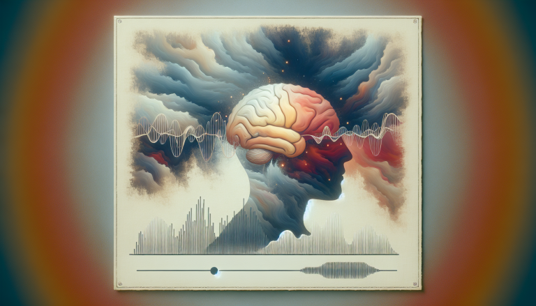
Tinnitus Linked to Altered Brain Blood Flow
Peer-Reviewed Research
A 2026 study from Capital Medical University in Beijing has identified a specific pattern of reduced blood flow in the brains of individuals with non-auditory tinnitus (NAT). The research, published in *Brain Imaging and Behavior*, links these perfusion deficits to impaired cerebral venous drainage, connecting a vascular condition directly to tinnitus symptoms and associated distress.
Key Takeaways
- Patients with non-auditory tinnitus (NAT) showed significantly reduced cerebral blood flow (CBF), particularly in the left hemisphere, compared to patients without tinnitus and healthy controls.
- The reduced blood flow was most pronounced in brain regions involved in attention, sensorimotor processing, and the default mode network.
- Lower CBF in NAT patients correlated with longer tinnitus duration, poorer sleep quality, and worse depression scores.
- The findings point to cerebral venous congestion (CVC) as a potential underlying vascular mechanism for certain types of tinnitus.
Linking Venous Congestion to Brain Perfusion
The research team, led by Lu Liu, Milan Jia, and senior authors Chen Zhou and Xunming Ji, focused on a condition known as cerebral venous congestion (CVC). CVC involves stenosis, or narrowing, in key veins like the internal jugular vein or cerebral venous sinuses. This narrowing impairs the brain’s ability to drain deoxygenated blood, which can secondarily reduce the inflow of fresh, oxygenated blood—a process measured as cerebral blood flow (CBF). While tinnitus is often considered an auditory system disorder, the “non-auditory” label here suggests the phantom sound may originate from broader neural or vascular dysfunction.
The study builds on a growing body of work examining vascular contributions to hearing disorders. For a broader look at how blood flow changes relate to tinnitus, see our article on Cerebral Blood Flow Changes in Tinnitus.
How the Study Measured Brain Blood Flow
The researchers conducted a cross-sectional study with 87 participants: 34 patients with CVC and NAT, 17 patients with CVC but without tinnitus (NAT-), and 36 healthy controls. To quantify blood flow, they used a non-invasive MRI technique called multi-delay pseudo-continuous arterial spin labeling (ASL). This method magnetically labels arterial blood water as an intrinsic tracer, allowing researchers to calculate CBF without contrast agents. The multi-delay aspect helped account for variations in arterial transit time, providing a more accurate picture of perfusion, especially in cases of possible vascular impairment.
CBF was measured across the whole brain and in 166 specific regions defined by the AAL3 atlas. The team then statistically compared perfusion between the three groups and analyzed correlations between CBF values and clinical data, including tinnitus duration, sleep quality (Pittsburgh Sleep Quality Index), and scores for anxiety, depression, and cognition.
Left-Sided Blood Flow Reductions Correlate with Symptoms
The results revealed a clear distinction. The NAT+ group exhibited significant CBF reductions compared to both the NAT- group and healthy controls. These reductions were most prominent in the left cerebral hemisphere. Specific brain regions with notably lower perfusion included the left insula, paracentral lobule, and precentral gyrus.
“These regions are not primary auditory areas,” the authors note. Instead, further analysis showed the affected areas belong to major functional networks: the attention network, sensorimotor network, default mode network (active at rest), and cerebellar network. This suggests the vascular impact of CVC disrupts integrated brain systems involved in monitoring internal and external states, movement planning, and self-referential thought—processes often altered in chronic tinnitus.
The clinical correlation data provided a direct link to patient experience. Within the NAT+ group, lower CBF was associated with a longer duration of tinnitus, poorer subjective sleep quality, and higher scores on depression scales. This strengthens the argument that the observed perfusion deficit is not just an incidental finding but is tied to the severity and burden of the condition.
The involvement of the cerebellum in this network analysis is particularly interesting, as this structure’s role in auditory and emotional processing is gaining recognition. You can explore this further in our resource on Cerebellar Insights for Tinnitus and Misophonia.
Implications for Understanding and Treating Tinnitus
This study offers a tangible, physiological mechanism for at least a subset of tinnitus cases. It moves the discussion beyond the cochlea and auditory cortex to include the brain’s vascular health. The pathophysiology proposed is that venous congestion creates a “backup” in the system, leading to sluggish cerebral perfusion that starves critical neural networks of optimal oxygen and nutrient supply, ultimately generating the phantom sound and its common comorbidities.
Practically, these findings could influence diagnostic pathways. For patients presenting with non-auditory tinnitus, especially with co-occurring sleep or mood complaints, assessment for potential venous congestion might become a consideration. This could involve specific MRI protocols to examine the cerebral veins and internal jugular veins.
Furthermore, the research opens a potential new avenue for treatment approaches. If venous congestion is a confirmed contributor, therapies aimed at improving venous outflow—which are currently explored for other neurological conditions—might be investigated for efficacy in relieving tinnitus and its associated symptoms. It underscores the importance of a holistic, brain-body approach to hearing health disorders.
The connection between vascular health and sensory processing also resonates with research on other sound sensitivity conditions. For a look at how brain responses differ in related disorders, read our analysis of Brain Responses to Sounds in Misophonia vs Hyperacusis.
Source Research
This article is based on the study “Cerebral blood flow alterations in non-auditory tinnitus: implications for cerebral venous congestion pathophysiology” by Liu L, Jia M, Li H, et al. (Brain Imaging Behav. 2026). The full paper is available via DOI: 10.1007/s11682-026-01144-8 or PMID: 41957332.
Evidence-based options: zinc picolinate, magnesium glycinate
Medical Disclaimer
This article is for informational purposes only and does not constitute medical advice. The research summaries presented here are based on published studies and should not be used as a substitute for professional medical consultation. Always consult a qualified healthcare provider before making any changes to your health regimen.
Peer-reviewed health research, simplified. Early access findings, clinical trial alerts & regulatory news — delivered weekly.
No spam. Unsubscribe anytime. Powered by Beehiiv.
Related Research
From Our Research Network
Exercise & metabolic fitnessSleep Science
Sleep & circadian healthPet Health
Veterinary scienceHealthspan Click
Longevity scienceBreathing Science
Respiratory healthMenopause Science
Hormonal health researchParent Science
Child development researchGut Health Science
Microbiome & digestive health
Part of the Evidence-Based Research Network